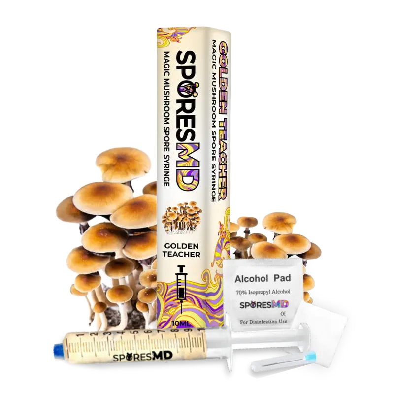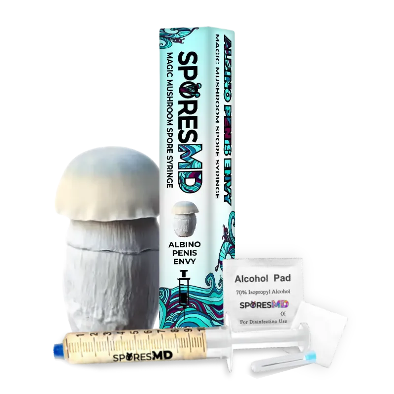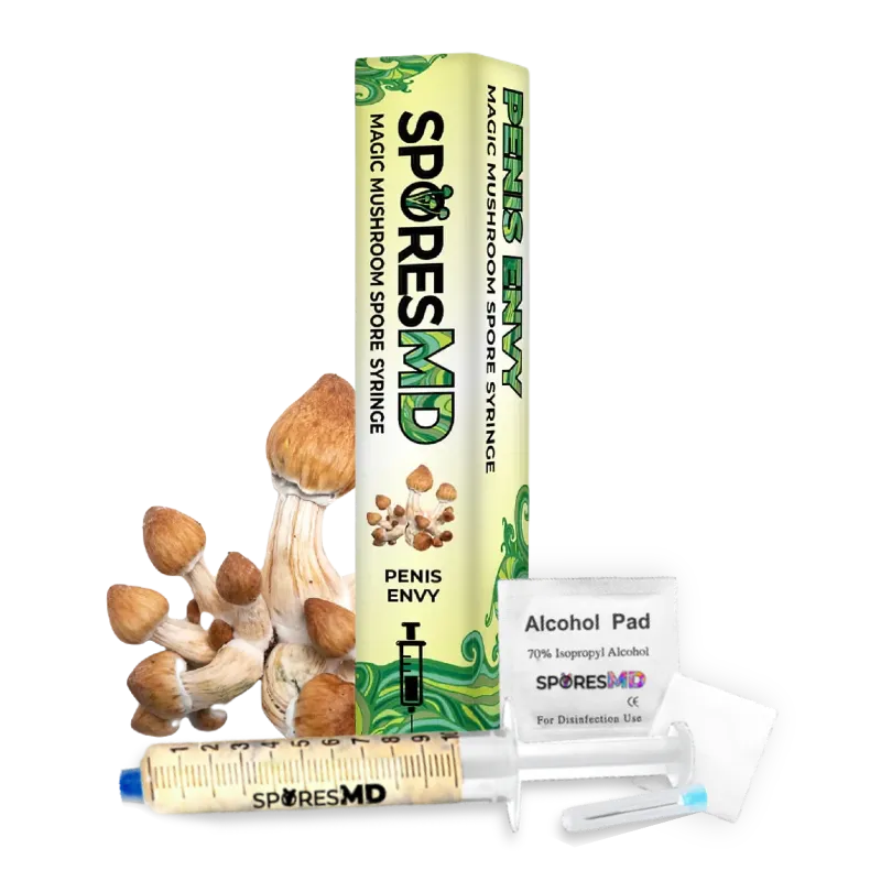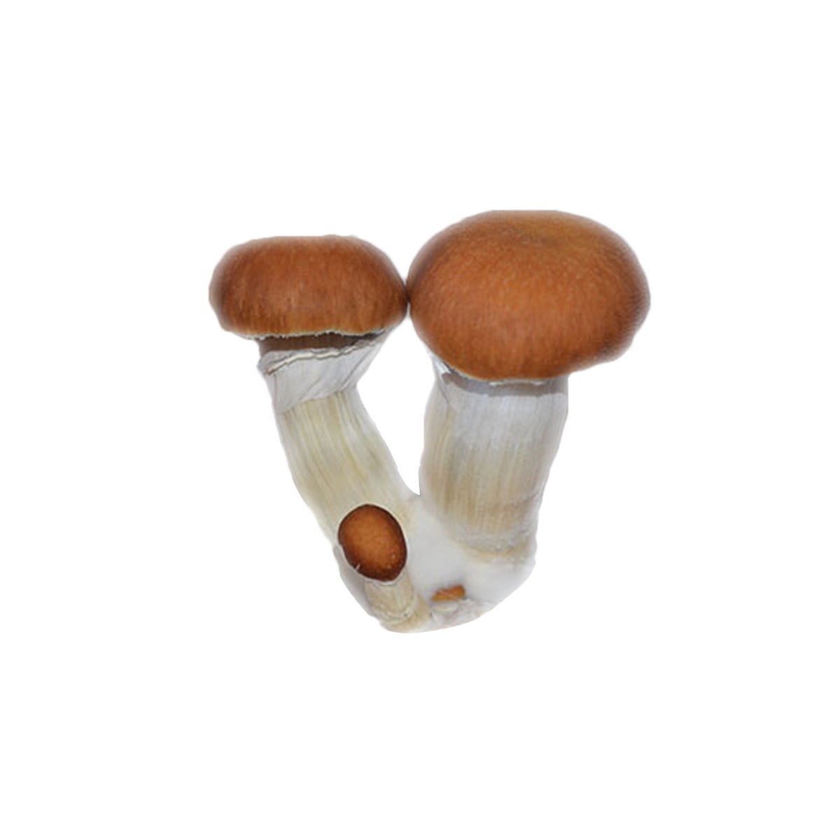
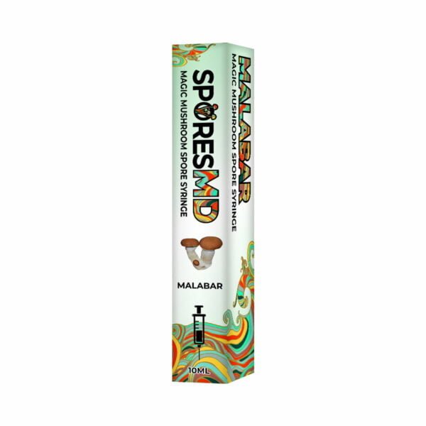
Malabar mushroom liquid culture from SporesMD delivers live Psilocybe cubensis mycelium in a sterile 10 mL syringe—optimized for 400x–1000x phase-contrast microscopy. Prepared in HEPA-filtered cleanroom environments with batch-level purity and viability validation.

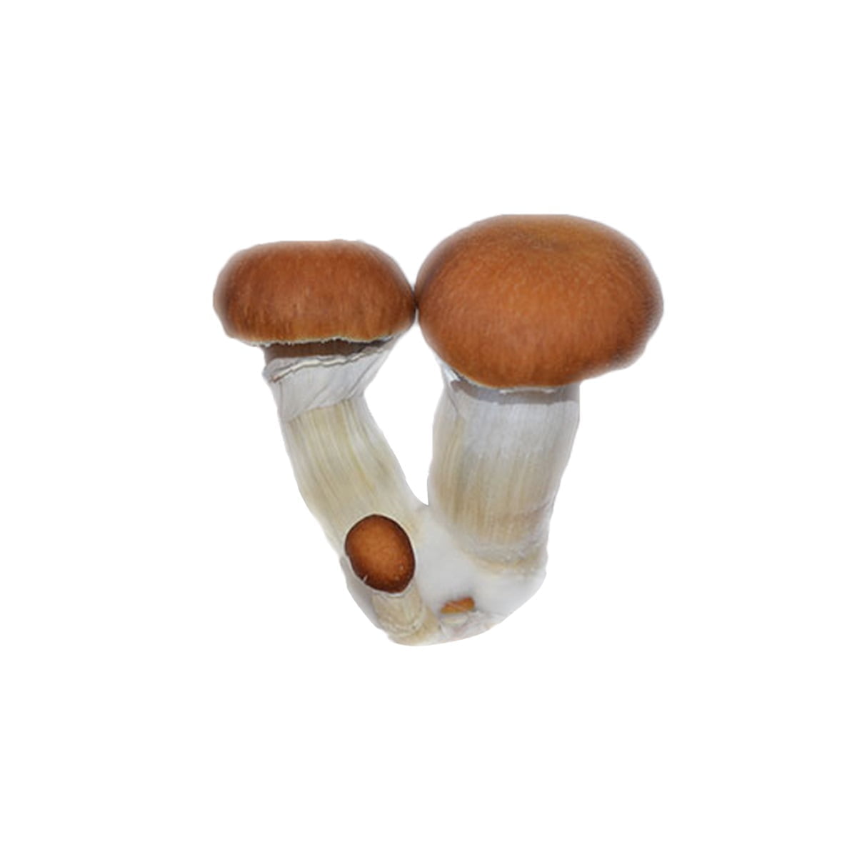
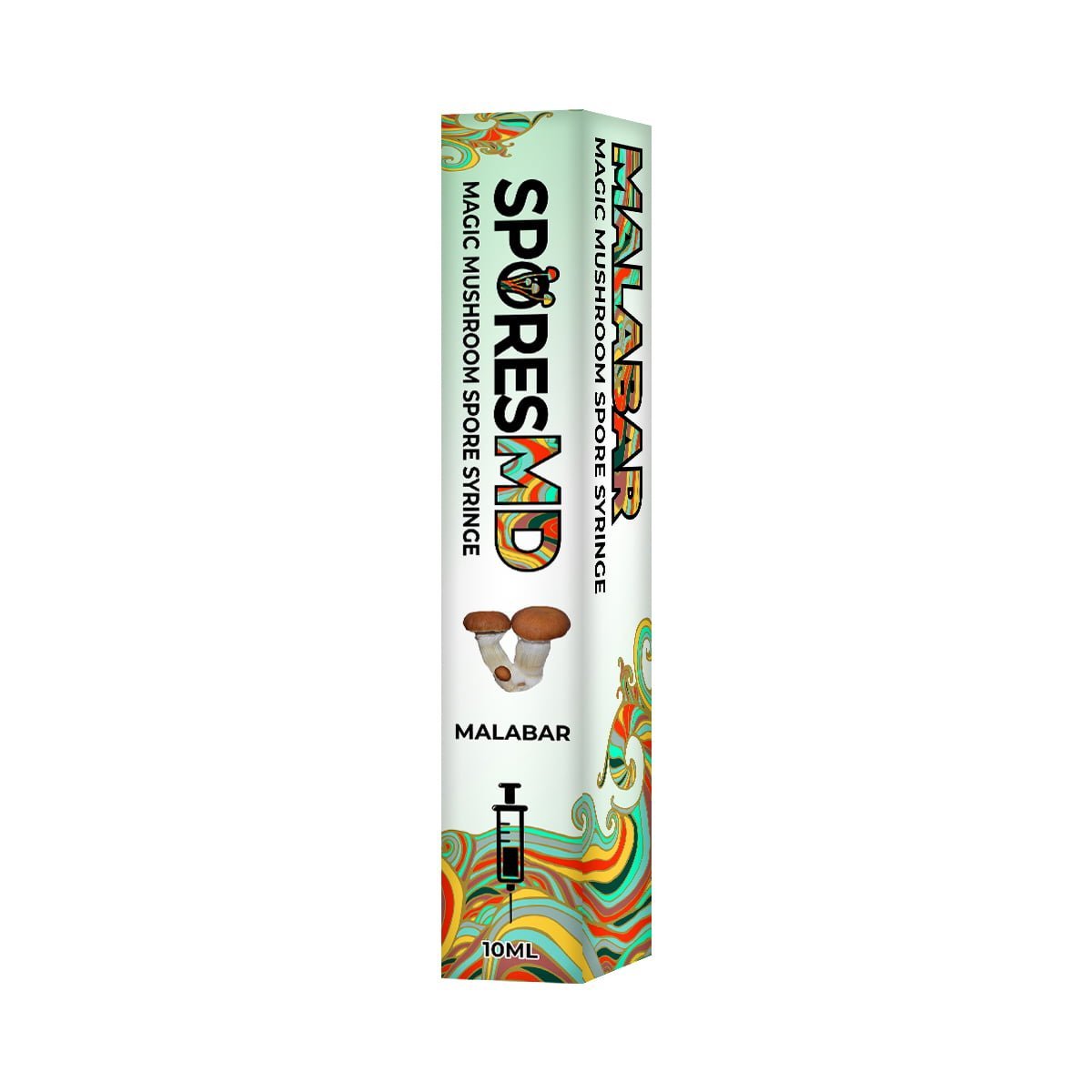
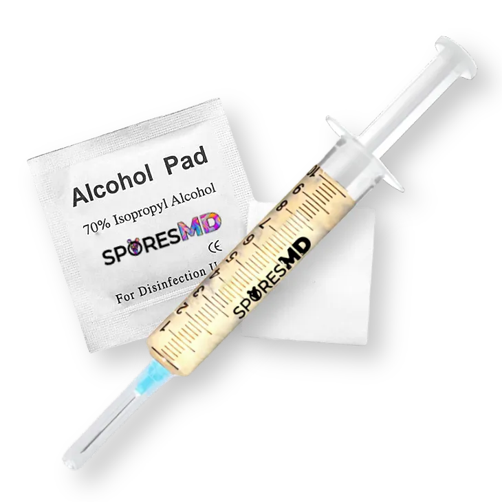

Malabar Mushroom Liquid Culture (10ml)
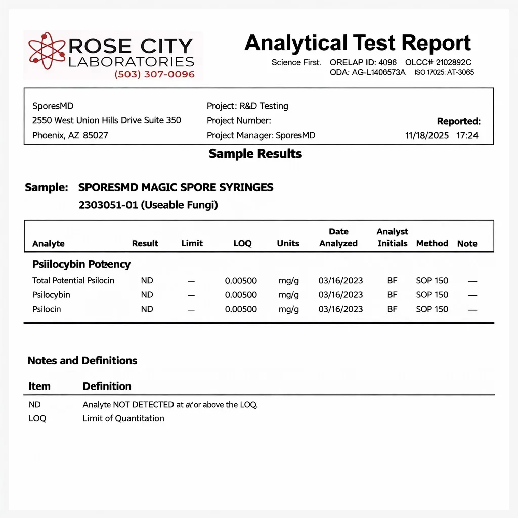
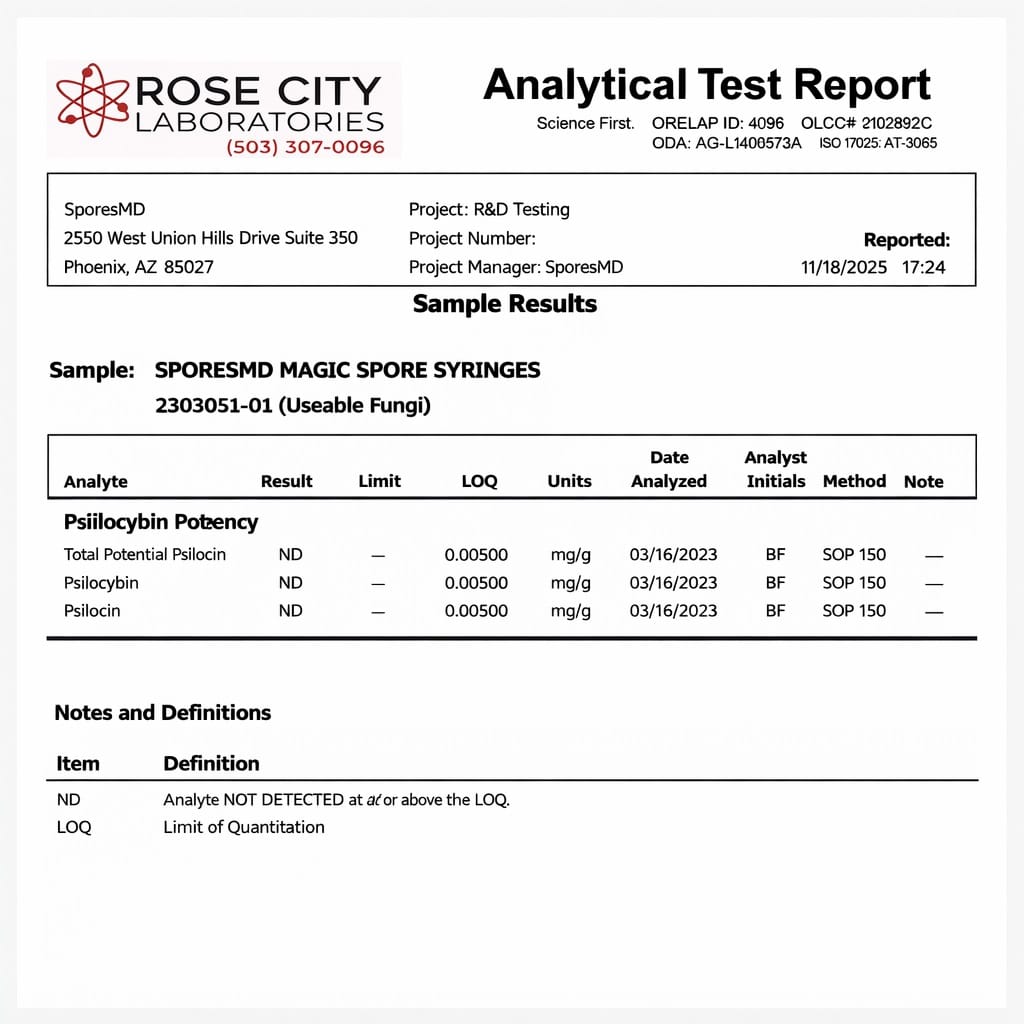
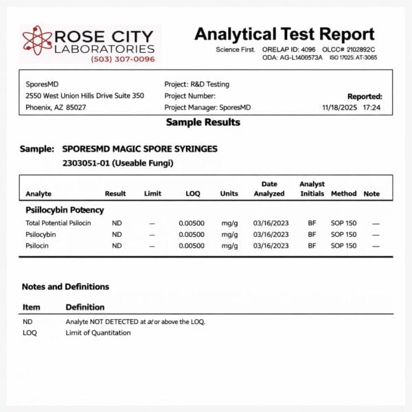
Immerse yourself in the realm of mycology with our Malabar Liquid Culture, contained in a 10mL syringe for precise academic research. Celebrated for its resilience and distinctive morphological traits, the Malabar strain is an excellent subject for studying fungal structure, genetics, and microscopy. Our commitment to excellence is reflected in each meticulously prepared batch, delivering a clean, reliable sample designed to support detailed laboratory and educational research. View Lab Report
| Feature | Specification |
| Volume | 10 mL sterile syringe |
| Contents | Live Malabar Psilocybe cubensis mycelium in nutrient solution |
| Preparation Environment | HEPA-filtered cleanroom |
| Recommended Magnification | 400x–1000x (brightfield or phase-contrast) |
| Storage Temperature | 2–8°C (35–46°F) |
| Shelf Life | Up to 12 months refrigerated |
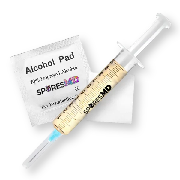
| Includes | Sterile needle, tamper-evident packaging |
| Price | $29.99 USD |
Live mycelium changes everything under the lens. Microscopy spore syringes show ungerminated structures. Liquid culture shows growth in motion—branching patterns, hyphal fusion, and colony formation. Each syringe—part of SporesMD’s liquid cultures for microscopy collection—contains actively developing Malabar mycelium suspended in nutrient-rich research solution, ready for slide preparation and detailed observation of hyphal networks, clamp connections, septa formation, and early-stage mycelial architecture.
What Are Malabar Mushrooms?
Malabar is a regional Psilocybe cubensis variety native to India’s tropical southwest coast. It thrives on dung-rich substrates in hot, monsoon-driven environments along the Arabian Sea—making it a compelling specimen for researchers studying how climate shapes cubensis morphology and sporulation. Those new to fungal reproductive biology can review what are mushroom spores for foundational context.
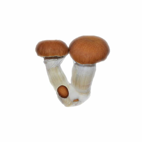
Under microscopy, Malabar produces thick, dense caps ranging from cinnamon to golden, and dark purple-to-black spore prints with sub-ellipsoid, smooth-walled mushroom spores. These characteristics offer consistent pigmentation across batches—ideal for side-by-side strain comparisons in controlled research settings.
Origin and Strain Identity
Malabar traces to India’s Malabar Coast—a historically significant trade region with dense vegetation, heavy monsoonal rainfall, and consistently high humidity. Early collections came from dung-rich coastal ecosystems in Kerala and adjacent states.
Within strain taxonomies, Malabar is classified as a regional cubensis variant, not a separate species. Spores measure approximately 11–13 µm × 7–9 µm with dense, random dispersion patterns visible on spore prints for microscopy. Notably, albino derivatives have been isolated from this lineage for experimental microscopy work.
Malabar Strain Characteristics
That geographic origin translates directly into observable morphology. Malabar produces large, dense fruiting bodies—caps ranging 50–75 mm in diameter—with golden to caramel coloration and strong white stems that bruise blue-green. Compared to other well-documented varieties like the golden teacher mushroom strain or penis envy mushroom, Malabar stands out for its dense cap tissue and slow-breaking veil.
| Feature | Detail |
| Cap diameter | 50–75 mm |
| Spore print color | Deep purplish-brown to black |
| Veil behavior | Slow-breaking, membranous annulus |
The partial veil remains attached longer than most cubensis lineages, which limits spore release timing—a distinctive trait under taxonomic study.
Spore Traits for Microscopy
Under magnification, Malabar spores present as sub-ellipsoid to ellipsoid basidiospores—smooth-walled with a prominent germ pore resolvable at higher numerical apertures. Researchers familiar with mushroom spore identification under microscopy will find Malabar specimens straightforward to characterize.
- Pigmentation: Uniform, dense cytoplasmic fill—strong contrast at 400x–1000x
- Surface: Smooth, non-ornamented walls
- Optimal setup: Phase-contrast or brightfield optics
In well-prepared cleanroom syringes, Malabar suspensions run dense. Thin slide preparations or dilution may be necessary for isolating individual spore morphology and imaging wall stratification. Researchers can also collect surface material using microscopy swabs for targeted sampling of specific structures.
SporesMD Malabar Liquid Culture Product Details
Each unit is prepared in HEPA-filtered cleanroom environments and validated via phase-contrast microscopy and agar-plate sterility checks. The syringe arrives in tamper-evident, individually sealed packaging with a sterile needle included. For researchers exploring where to buy mushroom spores from verified sources, the sporesmd homepage offers syringes, prints, swabs, and liquid cultures across dozens of strains.
Store at 2–8°C (35–46°F)—never freeze. Under proper refrigeration, viability holds up to 12 months—see spore syringe shelf life for detailed storage timelines. This product is sold strictly for microscopy, taxonomy, and educational research. Because psilocybin is a Schedule I controlled substance under U.S. federal law, legal status and restrictions vary by jurisdiction—researchers should review the legality of mushroom spores and are responsible for understanding and complying with all applicable laws.
Microscopy Usage & Handling Guidelines
Proper technique determines everything. Use aseptic methods throughout. Prepare slides inside a sanitized still-air box or laminar flow hood. Wipe all surfaces and tools with 70% isopropyl alcohol before opening any sealed applicator. For a complete walkthrough on preparation and observation, see how to use mushroom spores under the microscope.
| Step | Action |
| Resuspend | Shake syringe gently; use vibration for clumps |
| Dispense | Expel a sesame-seed-size droplet onto slide center |
| Coverslip | Lower at an angle—one edge first—to eliminate bubbles |
| Observe | Start low magnification, then move to 400x–1000x |
| Store | Refrigerate at 2–8°C (35–46°F); never freeze |
Label every slide with strain, date, and source for reproducible research records. Flame-sterilize needles until red-hot between uses and allow cooling before contacting solution. For long-term storage best practices, see how to store spore syringes.
FAQ: Malabar Shrooms vs Thai Mushroom Strains
How does Malabar compare to Thai-origin cubensis strains under the microscope?
Both are Psilocybe cubensis. Both produce dark purple-brown spores. But their lineage-specific traits create distinct microscopy focal points.
| Feature | Malabar (India) | Thai Strains (Thailand) |
| Spore Density on Prints | Lighter deposits—veil stays attached longer | Heavy, abundant spore drops |
| Spore Shape | Subellipsoid, thick-walled | Ellipsoid to subellipsoid, densely pigmented |
| Key Microscopy Focus | Veil persistence, annulus structure, basidial architecture | Germ pore visibility, print density, island vs. mainland variation |
| Available Formats (SporesMD) | Spore syringe + liquid culture | Spore syringe (e.g., Pink Buffalo) |
Malabar rewards researchers studying low-dispersal morphology and mycelial development. Thai lines excel at comparative spore-field analysis at 400x–1000x. Both are available alongside dozens of other varieties—browse the full product catalog for the complete research lineup.
For microscopy, taxonomy, and educational research only. Not for cultivation or ingestion.