


Spores MD
2550 West Union Hills Drive
Suite 350 – #9222
Phoenix, AZ 85027
Showing all 5 results
Microscopy spores provide access to the finer details of fungal biology, yet these details can only be seen clearly when the spores are prepared and handled with precision. Without proper preparation, the structures can appear unclear, and the distinctions between microscopy strains become difficult to identify.
When viewed through reliable microscopy samples, the spores display distinct shapes, surface textures, density patterns, and identifying characteristics that mark one strain from another. This level of visibility supports accurate comparison, detailed study, and a deeper understanding of fungal variation.
SporesMD offers microscopy-grade spores prepared specifically for research and educational viewing. Each sample is handled with careful attention to clarity and consistency so that the features present in the spores are visible under magnification. All microscopy spores from us are intended solely for research and observation and are not offered for cultivation or consumption. Explore our full microscopy spore selection today and find the strain that matches your research goals.
Understanding the purpose and qualities of microscopy spores creates a foundation for meaningful research and accurate examination.
Microscopy spores are individual fungal reproductive cells prepared for magnified observation. Each spore carries physical traits that reflect its lineage. Certain microscopy spores are particularly valued for study due to their consistent structural patterns. Viewed under magnification, these spores reveal fine details such as wall texture, surface smoothness, curvature, coloration variations, and differences in size or density.
Spore morphology helps researchers compare related strains and recognize subtle distinctions. With repeated observation, the eye becomes familiar with traits that define specific varieties. Observing spores does not require advanced laboratory tools, only appropriate slide preparation and a standard compound microscope.
The quality of microscopy spores directly affects how clear, accurate, and useful the viewing experience will be. When spores are not prepared or handled correctly, the ability to identify and compare structures becomes limited. Reliable preparation and strain verification make it possible to observe the true characteristics of each sample without interference.
High-quality spores provide clarity and structure that support both learning and precise comparison. This consistency is especially valuable when developing familiarity with strain differences under magnification.
Purchasing microscopy spores from a reliable source directly affects the efficiency and clarity of research sessions. SporesMD provides several benefits that support precise viewing and accurate comparison.
Spores are suspended in sterile solution and packaged in environments designed to minimize contamination. This helps ensure visible clarity during examination. Clean preparation reduces the risk of bacterial interference, clouding, or discoloration that obstructs spore structure.
Each strain sold by SporesMD has a confirmed lineage. This accuracy allows meaningful comparison across multiple strains without uncertainty. Researchers who document observations benefit from knowing the visual differences they see represent real genetic variation rather than mislabeled samples.
Each syringe contains a consistent spore concentration. Balanced density supports the quick location of spores once placed on a slide. Sparse samples waste time and reduce observation quality. SporesMD ensures samples are dense enough for efficient and repeatable study.
Orders are packaged securely to protect sterility. Shipping is carried out discreetly to maintain privacy and ensure that the materials arrive in usable condition.
These combined benefits allow researchers to focus on observation rather than troubleshooting preparation issues. Browse our microscopy spores collection now and begin building your research library with consistency.
The strains available from SporesMD each provide their own distinctive visual characteristics. This creates options for both new observers and experienced analysts.
B+ spores typically display elliptical shapes and consistent outlines. Their symmetry makes them helpful for developing familiarity with foundational spore structure. The stable size distribution observed in B+ spores supports foundational training in identification.
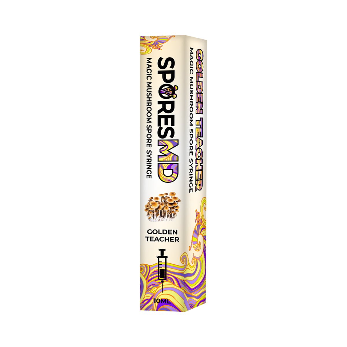
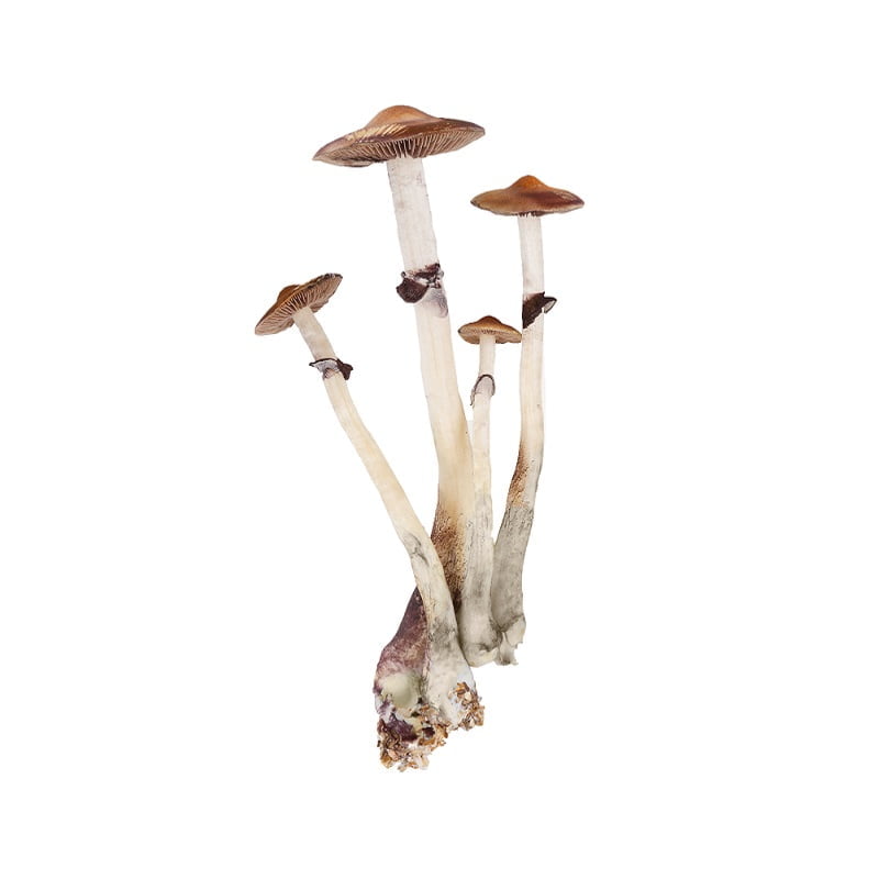
Golden Teacher spores often show slight variability in size and smooth surface textures. Observing Golden Teacher can help users understand how small fluctuations in dimensions affect the overall visual profile under magnification.
Jedi Mind spores exhibit surface and shape features that stand out from more standardized varieties. This strain can be useful for observers who want to expand their comparison sets and recognize structural variations with confidence.
Pink Buffalo spores are valued for their clean outlines and consistent form. Their structure can offer clarity during early microscopy work, while also providing subtle characteristics for comparison at higher magnification.
Tidal Wave is a hybrid strain. The spores often show unique density patterns and small visual distinctions from traditional varieties. Researchers often include Tidal Wave in comparative analysis due to its distinct hybrid lineage.
These strains provide a spectrum of morphological features that support both introductory and advanced microscopy research.
Viewing microscopy spores requires deliberate slide preparation and consistent control over the microscope settings. The clarity of the observation depends heavily on how the sample is placed, how the cover slip is positioned, and how the magnification is adjusted. When handled correctly, the spores will appear sharply defined, allowing for easy identification of shape, texture, and density patterns that distinguish microscopy strains.
A careful approach is what allows the microscopic features of the spores to show clearly. The shape of each spore, the thickness of the outer wall, the natural tint within the cell, and the distribution of spores across the field all become easier to evaluate when magnification is increased gradually and lighting is adjusted in small steps. Over time, these repeated observations lead to a deeper familiarity with how microscopy varieties differ from one another.
Consistent technique also contributes to reliable comparison. When the same slide preparation method is repeated across multiple strains, the differences between them stand out more clearly. This makes it easier not only to study individual spores but also to recognize repeating visual patterns across entire varieties. Explore our selection of microscopy spores that offer strong visibility and preserved structure for clear viewing.
Studying microscopy spores supports learning, research, and hands-on scientific exploration across multiple groups and skill levels.
Students develop an understanding of fungal reproduction and structural diversity by observing spores. Microscopy provides a direct view of morphological traits, supporting classroom learning with real visual reference points.
Hobbyists use microscopy spores to study lineage variation and gain experience in recognizing strain-specific characteristics. This practice encourages independent research and personal growth in fungal knowledge.
Researchers studying fungi benefit from clear and verified spore samples. Reliable strain identity supports structured documentation and comparative analysis, which is essential in academic or reference-based research settings.
Microscopy spores provide hands-on learning material for lessons in genetics, morphology, and microbial observation. The visual clarity and predictable structure make them suitable for teaching foundational microscope skills.
In each setting, microscopy spores support the development of analytical skills and deeper familiarity with fungal biology.
Microscopy spores from SporesMD are for research and educational viewing only. They are not intended for cultivation. Users are responsible for understanding and following all local regulations.
Microscopy spores provide a direct view into the microscopic structure of fungi. Each strain carries its own distinctive characteristics, and observing these variations supports research, comparison, and learning. Reliable spores allow for clear examination without interference or uncertainty.
SporesMD provides microscopy spores that are cleanly prepared, accurately identified, and consistently dense, allowing observers to focus on the visual and structural details that make microscopy rewarding.
Select your microscopy spores from SporesMD to begin your next session of research and observation.
2550 West Union Hills Drive
Suite 350 – #9222
Phoenix, AZ 85027
Disclaimer: SporesMD specializes in providing spore specimens exclusively for microscopy and educational use. Our products are strictly not intended for cultivation purposes, and we firmly oppose any such use. Customers are responsible for ensuring compliance with all relevant laws and regulations regarding spore possession and use. SporesMD accepts no liability for damages, injuries, or legal issues resulting from the use or misuse of our products. Our commitment is to the quality of our spore specimens for scientific and educational study, without warranties except as mandated by law.
© 2026 SporesMD. All Rights Reserved.